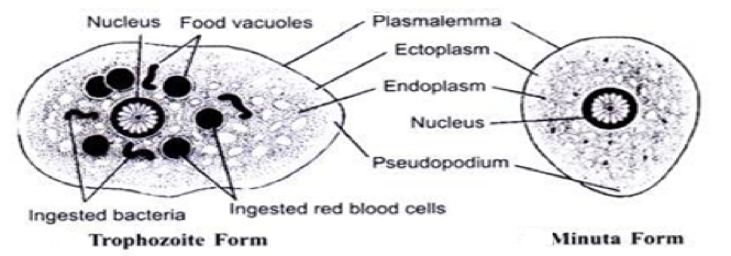
Entamoeba histolytica

Morphological Characteristics
The flow of the ectoplasm and endoplasm allows for motility. This parasite contains a nucleus and food vacuoles. The minuta form is the stage in between the cyst and trophozoite stage.
Identification Methods
Microscopic investigation of stool samples is used to detect cysts and trophozoites.
Life Cycle
- Cysts and tophozoites are passed in feces of humans
- Infection generally occurs by formed cysts which are ingested through food, water, or hands
- Excystation (exit from cyst stage) occurs in small intestine
- Trophozoites are released and migrate to large intestine
- Trophozoites multiply by binary fission. Cysts are produced.
HOST INFORMATION
- Primarily human hosts (no intermediates)
- People are more at risk in areas which have poor sanitation since this parasite is transmitted via faecal-oral route
- Is also common in men who have sex with men
- Can often be carried with no symptoms, making spread easy
- Antibiotics can be used to treat this parasitic infection
GEOGRAPHIC DISTRIBUTION
Entamoeba histolytica is distributed worldwide. The majority of infections occur in developing countries due to poor sanitation.
SOURCES
https://www.cdc.gov/parasites/amebiasis/pathogen.html
https://www.ncbi.nlm.nih.gov/pmc/articles/PMC3378717/
https://www.waterpathogens.org/book/entamoeba-histolytica
https://www.health.ny.gov/diseases/communicable/amebiasis/fact_sheet.htm