Zaire ebolavirus
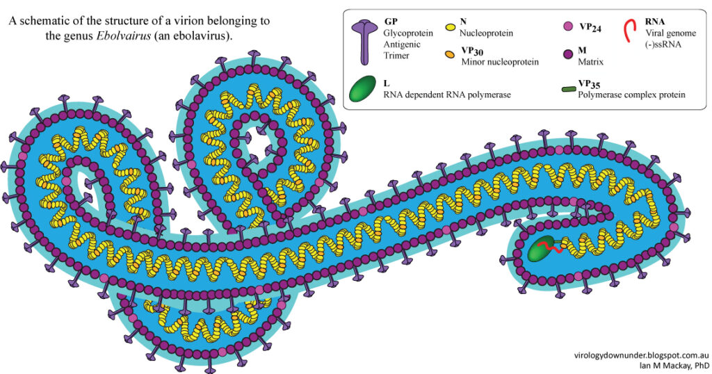
Morphological Characteristics
Ebola virus has a tubular structure which is surrounded by a membrane with proteins on the outer membrane. The RNA resides in the matrix in cylindrical virions.
Identification Methods
PCR is used to detect virus particles from blood samples.
Life Cycle
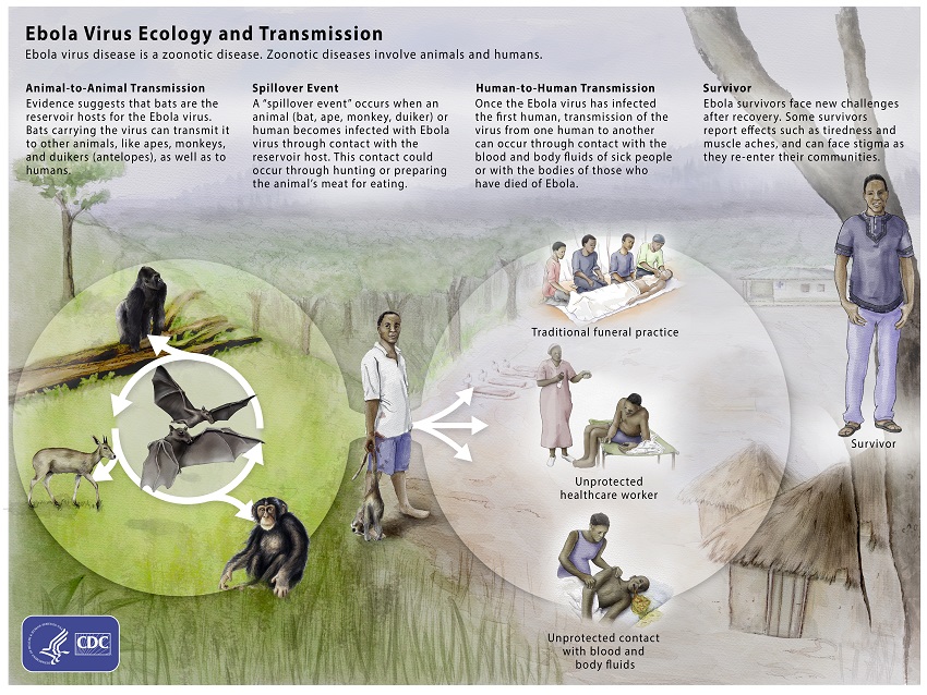
-resevoir host is bats
-humans can get it from contact with an animal which is infected and then can spread it
-Entry: Virus binds to receptors of cells and enters through macropinocytosis
*macropinocytosis: inward folding by the cell surface so that membrane forms macropinosomes carrying the viral RNA
-Uncoating and fusion: The protein NCP1 induces fusion of EBOV and membrane of macropinosome so that the genome is now in the cell cytoplasm
-Transcription and replication: The EBOV RNA is transcribed to create viral proteins called RNP
-Assembly: The viral proteins assemble new ebolavirus
-Budding: Occurs at cell membrane. The new virus becomes infective so that replication can continue and is released either outside of the cell or continues replication within the same cell.

HOST INFORMATION
- reservoir hosts: bats
- Other animal hosts: apes, monkeys, antelopes, humans
- Spillover from animal to human through hunting
- Human to human transmission: occurs through contact with blood/ body fluids of an infected person or by someone who has died of Ebola
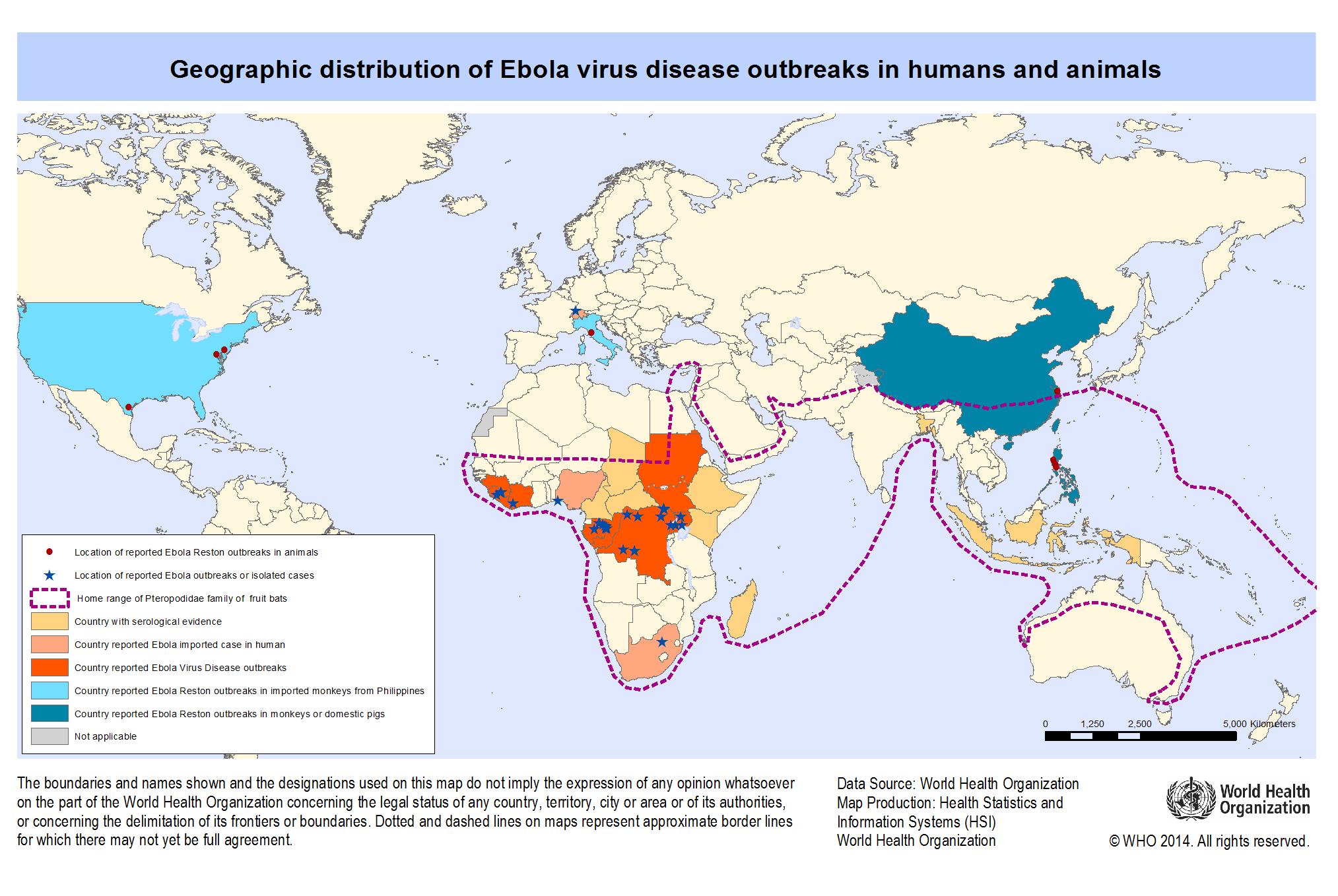
Geographic Distribution
The ebola epidemic lasted from 2014 to 2016. This map shows the zones which were most affected during the epidemic. The purple dotted line shows the range of the reservoir host.
SOURCES
https://doi.org/10.18632/oncotarget.18498
https://www.youtube.com/watch?v=BHQUp-R0q9U
https://www.cdc.gov/vhf/ebola/about.html
https://www.cdc.gov/vhf/ebola/history/2014-2016-outbreak/index.html
https://microbewiki.kenyon.edu/index.php/Ebola_Virus_Disease